
Cellular Signaling
Nearly 150 years ago, Charles Darwin reported phototropism of the coleoptile in plants1. Since then, scientific understanding of cellular signaling processes has exploded. Cell signaling is the foundation of nearly all biological processes, from cell division, differentiation, growth, and nutrient cycling to cell death2. Specialized processes like neurotransmission, pathogen-sensing, and antigen-presentation are also controlled by cell signaling2.
The Players3-22
Several physical components make up the “players” of cell signaling.
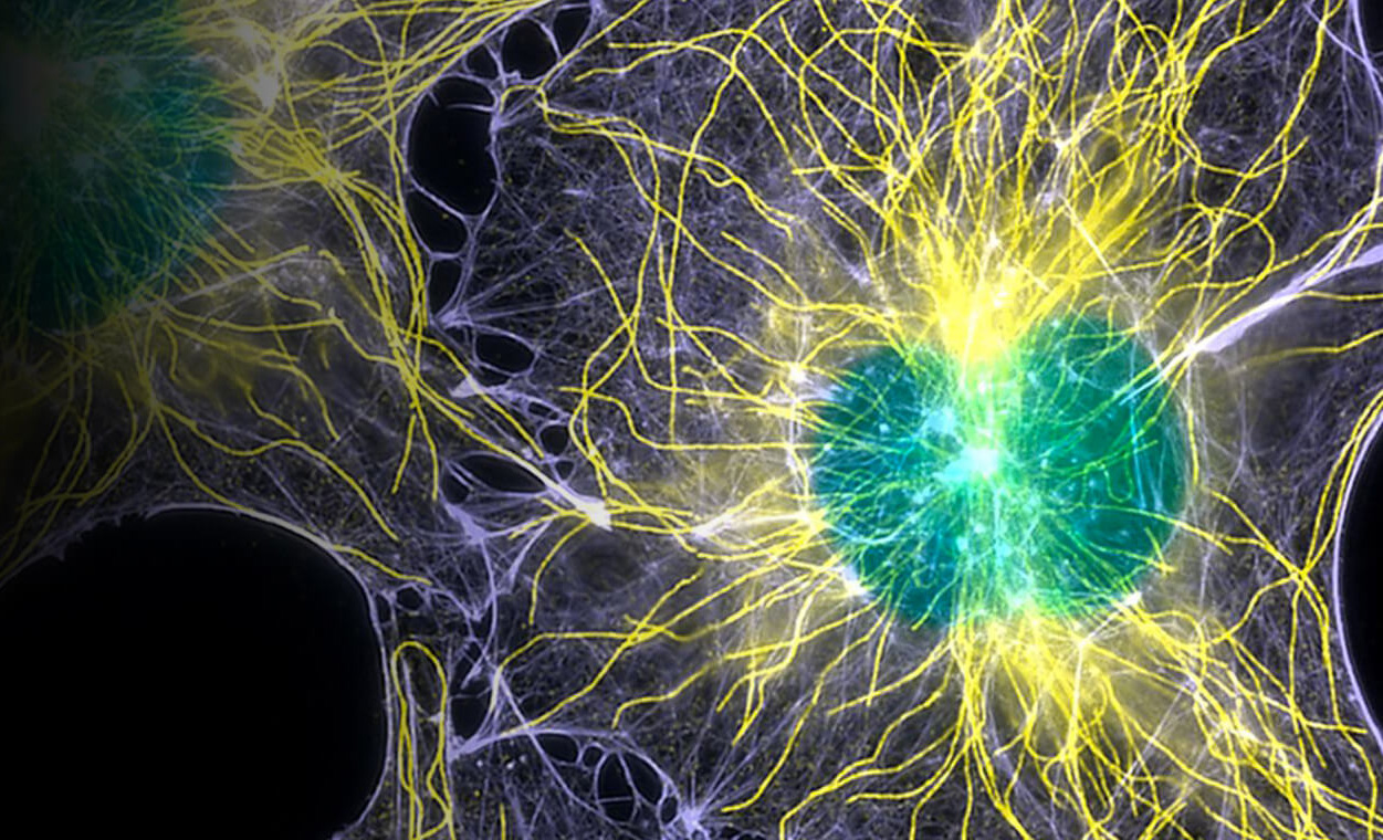
Cell Membranes3-4
The first player is the cell membrane, the phospholipid bilayer barrier across which the signal is transmitted. The barrier is not just a passive participant, either. Research shows that the types and ratios of lipids making up the membrane affect several aspects of cell signaling.
The membrane composition affects the way membrane receptor proteins interact with and embed in the membrane, as well as how permeable the membrane is to ligands with intracellular receptors. The membrane composition also affects the sensitivity and kinetics of some ligand-receptor interactions. Membrane lipids can directly associate with membrane receptors or act as second messengers.
Finally, the three-dimensional structure of the membrane is now known to include lipid rafts, which can act as an assembly scaffold for membrane proteins. Extrusions and invaginations also contribute to the stability, exposure, and removal of membrane receptors, as well as endocytosis or exocytosis of ligands.
Ligands5-7
The next players are ligands, small molecules that transmit information from a source to the receiving cell. Ligands tend to bind only with specific receptors in a “lock-and-key” or “hand-in-glove” type model5. The structural specificity, chemical affinity, and thermodynamic kinetics of ligand binding all influence signaling outcomes and are frequent targets of guided drug design6-7.
- Proteins, peptides, amino acids, and amino acid derivatives (ex: growth factors, neurotransmitters)
- Lipids (ex: steroids)
- Carbohydrates (ex: β-glucan)
- Small inorganic molecules (ex: nitric oxide)
Biofluids8
Human body mass is 50-75% water. Water-based biofluids are the medium through which ligands are transmitted between the source and the receptor(s).
- Intracellular fluid/cytosol (about 60% of the body’s water content)
- Extracellular fluid (about 40% of the body’s water content)
- Interstitial Fluid (about 30% of the body’s water content)
- Blood plasma (about 8% of the body’s water content)
- Others (about 2% of the body’s water content): Lymphatic fluid, cerebrospinal fluid, synovial fluid, pleural fluid, pericardial fluid, peritoneal fluid, and aqueous humor
Receptors9-21
Receptors are proteins that are activated by ligand binding and undergo conformational changes to initiate cell signaling cascades. Receptors are divided into two major classes: intracellular and cell-surface.
Intracellular Receptors9-14
Intracellular receptors respond to hydrophobic and lipid-soluble ligands that are capable of crossing the plasma membrane and binding to receptors in the cell interior.
Nuclear receptors typically act by directly binding to DNA sequences called hormone response elements and then modulating transcription9. The largest group of nuclear receptors are steroid hormone receptors10. Nuclear receptors include two main types:
- Type I receptors exist in the cytoplasm anchored by chaperone proteins. The binding of the ligand displaces the chaperone, allowing the receptor to homodimerize and expose the nuclear localization sequence. The homodimerized receptor-ligand complex then enters the nucleus and associates with transcriptional coactivators that facilitate binding to and activation of target genes.
- Type II receptors, in contrast, originate in the nucleus bound to their specific DNA response elements, where they exert a repressive influence via interaction with co-repressors. Ligand binding causes the corepressors to be replaced by coactivators that facilitate the activation of target genes.
Cytoplasmic and organellar receptors bind hydrophobic ligands, which pass through the cell membrane. Some examples include receptors for glutamate, thyroid hormone, estrogen, and androgens on the mitochondrial membrane11-12; sigma receptors on the ER membrane13; and the mannose-6-phosphate receptor on Golgi and lysosomal membranes14.
Cell-Surface Receptors15-21
Cell-surface receptors are anchored to or embedded in the plasma membrane and bind ligands from the extracellular space. The three main classes of cell surface receptors are ion channel-linked receptors, G protein-coupled receptors (GPCRs), and enzyme-linked receptors.
G-protein-coupled receptors (GPCRs)15 are the largest family of transmembrane signaling molecules in the human genome. These receptors are composed of a single polypeptide chain with a characteristic 7-α-helix transmembrane spanning domain. Ligand binding induces conformation change of the receptor, which activates intracellular signaling via a heterotrimeric guanosine triphosphate-binding protein (G-protein). In addition to the hundreds of known GPCRs, the human genome encodes at least 20 different α subunits, 6 β subunits, and 12 γ subunits of the G-protein. These channels are involved in an enormous variety of cell signaling processes and have been extensively studied. Over 370 structures of 70 unique GPCRs have been published, and about 35% of pharmaceuticals currently in use act on these receptors or their ligands16-17.
Ion channel receptors are ligand-binding receptors with intrinsic ion channel activity, also known as ligand-gated ion channels (LGICs). The largest group of ion channel receptors is the fast-acting ligand-gated ion channel superfamily18. This superfamily includes nicotinic ACh, 5-HT3, GABAA, and glycine receptors—all of the primary neurotransmitter receptors active in the nervous system.
Another more heterogeneous group of ion channel receptors are the acid-responsive ion channels, including the acid-sensing ion channels (ASICs), ionotropic purinoceptors (P2X) channels, inward rectifier K+ channels, voltage-activated K+ channels, L-type Ca2+ channels, hyperpolarization-activated cyclic nucleotide-gated channels, gap junction channels, and Cl–channels19. Several members of the transient receptor potential (TRP) channel superfamily are also considered ion channel receptors20.
Enzyme-linked receptors21 bind extracellular ligands, which induce a conformational change in the intracellular portion of the protein. This group of receptors is defined by either having an intrinsic enzymatic activity of the intracellular domain or having an intracellular domain that is directly associated with an enzyme. They have been categorized into 6 classes thus far:
- Receptor tyrosine kinases (RTKs) — Phosphorylate-specific tyrosines on intracellular signaling proteins
- Tyrosine-kinase-associated receptors — Associate with intracellular tyrosine kinases
- Receptor-like tyrosine phosphatases — Remove phosphate groups from tyrosines of specific intracellular signaling proteins
*note their status as receptors is unclear since their ligands have yet to be identified*
- Receptor serine/threonine kinases — Phosphorylate-specific serines or threonines on gene regulatory proteins to modulate transcription
- Receptor guanylyl cyclases — Directly catalyze the production of cyclic GMP in the cytosol
- Histidine-kinase-associated receptors — Associate with an intracellular kinase that autophosphorylates its histidine and then transfers the phosphate to another intracellular signaling protein
Second Messengers22
Second messengers are small molecules that transmit the signal from the ligand-receptor complex to an intracellular target. Classes of second messengers include:
- Cyclic nucleotides (ex: cAMP) — Signal within the cytosol
- Lipids (ex: IP3) — Signal within cell membranes
- Ions (ex: Ca2+) — Signal within and between cellular compartments
- Gases and free radicals (ex: Nitric Oxide) — Signal within and between cells
The Processes23-24,15
Cell signaling can be grouped into four main mechanisms and consists of three main steps.
Four Main Mechanisms23
- Paracrine — Signaling from one cell to nearby cells (ex: neurotransmitters)
- Autocrine — Signaling from one cell back to itself
- Endocrine — Signaling from specialized secretory cells through the bloodstream to distant cells
- Juxtracrine — Signaling between cells in direct contact
- Gap junctions — Allow molecules to pass between the cytosol of two adjoining cells (ex: in the lens of the eye)
- Surface-marker-receptor binding (ex: immune system)
Three Main Phases24,15
- Synthesis and release of signaling ligands
- Signal reception — Ligands bind to receptors
- Signal transduction — Receptors initiate an intracellular signal cascade
- The cascades can converge, diverge, or engage in crosstalk
- Many cascades employ second messengers
Common Pathways in Human Health and Disease25-41
- Since their discovery in the late 1800s, cell signaling pathways have been studied in pre-clinical research. After identifying the receptors and ligands from these pathways, research changed to focus on targeting these components for the treatment of human diseases. Some common signaling pathways being studied in vitro and in vivo for pre-clinical research and clinical trials for human diseases are:Notch25-26
- Hedgehog27-28
- TGF-β29-30
- Wnt/β-Catenin31
- Ras/Raf/MAPK32
- JAK/STAT33-34
- NF-kB35-36
- TNF37
- AKT/PI3K/mTOR38-39
- p5340-41
Currently, bench-to-bedside translational research has targeted the components of cell signaling pathways to treat cancer (all of the listed pathways), neurodegenerative diseases (Wnt/β-Catenin, JAK/STAT, TNF, AKT/PI3K/mTOR, and p53 pathways), autoimmune conditions (JAK/STAT, NF-kB, and TNF pathways), cardiovascular diseases (Notch pathway), osteoarthritis (Hedgehog pathway), fibrosis (TGF-β pathway), metabolic disorders (Wnt/β-Catenin pathway), and more25-41. The JAK/STAT and NF-kB pathways have even been targeted during the ongoing pandemic for their inflammatory role in SARS-CoV2 infection.
Here at Leo Corps, Inc., we are raising the bar for human disease research and discovery. We are dedicated to providing the highest quality products, including primary cells and in vitro and in vivo reagents for biomedical research, drug development, and translational investigations. Contact us today to find out how we can support your research!
References:
- Darwin C, Darwin FE. The ‘Power of movement in plants.’--1880. Published online 1888.
- Nair A, Chauhan P, Saha B, Kubatzky KF. Conceptual Evolution of Cell Signaling. Int J Mol Sci. 2019;20(13):3292. Published 2019 Jul 4. doi:10.3390/ijms20133292
- Cheng X, Smith JC. Biological Membrane Organization and Cellular Signaling. Chem Rev. 2019;119(9):5849-5880. doi:10.1021/acs.chemrev.8b00439
- Sunshine H, Iruela-Arispe ML. Membrane lipids and cell signaling. Curr Opin Lipidol. 2017;28(5):408-413. doi:10.1097/MOL.0000000000000443
- Koshland Jr DE. The key–lock theory and the induced fit theory. Angewandte Chemie International Edition in English. 1995;33(23‐24):2375-2378.
- Borisov DV, Veselovsky AV. Kinetika sviazyvaniia liganda s retseptorom v razrabotke lekarstv [Ligand-receptor binding kinetics in drug design]. Biomed Khim. 2020;66(1):42-53. doi:10.18097/PBMC20206601042
- Sriramulu DK, Lee SG. Combinatorial Effect of Ligand and Ligand-Binding Site Hydrophobicities on Binding Affinity. J Chem Inf Model. 2020;60(3):1678-1684. doi:10.1021/acs.jcim.9b01143
- Betts JG, Young KA, Wise JA, et al. Fluid, Electrolyte, and Acid-Base Balance. In: Anatomy and Physiology. OpenStax; 2013.
- Sever R, Glass CK. Signaling by nuclear receptors. Cold Spring Harb Perspect Biol. 2013;5(3):a016709. Published 2013 Mar 1. doi:10.1101/cshperspect.a016709
- Cheskis BJ. Regulation of cell signalling cascades by steroid hormones [published correction appears in J Cell Biochem. 2004 Sep 1;93(1):214]. J Cell Biochem. 2004;93(1):20-27. doi:10.1002/jcb.20180
- Psarra AM, Sekeris CE. Steroid and thyroid hormone receptors in mitochondria. IUBMB Life. 2008;60(4):210-223. doi:10.1002/iub.37
- Gough NR. Neuroprotective mitochondrial glutamate receptors. Science Signaling. 2012;5(247):ec272-ec272.
- Tesei A, Cortesi M, Zamagni A, et al. Sigma Receptors as Endoplasmic Reticulum Stress "Gatekeepers" and their Modulators as Emerging New Weapons in the Fight Against Cancer. Front Pharmacol. 2018;9:711. Published 2018 Jul 10. doi:10.3389/fphar.2018.00711
- Brown WJ, Farquhar MG. The mannose-6-phosphate receptor for lysosomal enzymes is concentrated in cis Golgi cisternae. Cell. 1984;36(2):295-307.
- Cooper GM. The Cell: A Molecular Approach. 2nd edition. Sunderland (MA): Sinauer Associates; 2000. Functions of Cell Surface Receptors. Available from: https://www.ncbi.nlm.nih.gov/books/NBK9866/
- Congreve M, de Graaf C, Swain NA, Tate CG. Impact of GPCR Structures on Drug Discovery. Cell. 2020;181(1):81-91. doi:10.1016/j.cell.2020.03.003
- Hauser AS, Attwood MM, Rask-Andersen M, Schiöth HB, Gloriam DE. Trends in GPCR drug discovery: new agents, targets and indications. Nat Rev Drug Discov. 2017;16(12):829-842. doi:10.1038/nrd.2017.178
- Ortells MO, Lunt GG. Evolutionary history of the ligand-gated ion-channel superfamily of receptors. Trends Neurosci. 1995;18(3):121-127. doi:10.1016/0166-2236(95)93887-4
- Holzer P. Acid-sensitive ion channels and receptors. Handb Exp Pharmacol. 2009;(194):283-332. doi:10.1007/978-3-540-79090-7_9
- Moran MM. TRP Channels as Potential Drug Targets. Annu Rev Pharmacol Toxicol. 2018;58:309-330. doi:10.1146/annurev-pharmtox-010617-052832
- Alberts B, Johnson A, Lewis J, et al. Molecular Biology of the Cell. 4th edition. New York: Garland Science; 2002. Signaling through Enzyme-Linked Cell-Surface Receptors. Available from: https://www.ncbi.nlm.nih.gov/books/NBK26822/
- Newton AC, Bootman MD, Scott JD. Second Messengers. Cold Spring Harb Perspect Biol. 2016;8(8):a005926. Published 2016 Aug 1. doi:10.1101/cshperspect.a005926
- Rye C, Wise R, Jurukovski V, DeSaix J, Choi J, Avissar Y. Cell Communication. In: Biology. OpenStax; 2016.
- Holz RW, Fisher SK. Cellular Signaling Mechanisms. In: Siegel GJ, Agranoff BW, Albers RW, et al., editors. Basic Neurochemistry: Molecular, Cellular and Medical Aspects. 6th edition. Philadelphia: Lippincott-Raven; 1999. Available from: https://www.ncbi.nlm.nih.gov/books/NBK28213/
- Majumder S, Crabtree JS, Golde TE, Minter LM, Osborne BA, Miele L. Targeting Notch in oncology: the path forward. Nat Rev Drug Discov. 2021;20(2):125-144. doi:10.1038/s41573-020-00091-3
- Aquila G, Kostina A, Vieceli Dalla Sega F, et al. The Notch pathway: a novel therapeutic target for cardiovascular diseases?. Expert Opin Ther Targets. 2019;23(8):695-710. doi:10.1080/14728222.2019.1641198
- Salaritabar A, Berindan-Neagoe I, Darvish B, et al. Targeting Hedgehog signaling pathway: Paving the road for cancer therapy. Pharmacol Res. 2019;141:466-480. doi:10.1016/j.phrs.2019.01.014
- Xiao WF, Li YS, Deng A, Yang YT, He M. Functional role of hedgehog pathway in osteoarthritis. Cell Biochem Funct. 2020;38(2):122-129. doi:10.1002/cbf.3448
- Liu S, Ren J, Ten Dijke P. Targeting TGFβ signal transduction for cancer therapy. Signal Transduct Target Ther. 2021;6(1):8. Published 2021 Jan 8. doi:10.1038/s41392-020-00436-9
- Lodyga M, Hinz B. TGF-β1 - A truly transforming growth factor in fibrosis and immunity. Semin Cell Dev Biol. 2020;101:123-139. doi:10.1016/j.semcdb.2019.12.010
- Ng LF, Kaur P, Bunnag N, et al. WNT Signaling in Disease. Cells. 2019;8(8):826. Published 2019 Aug 3. doi:10.3390/cells8080826
- Song Y, Bi Z, Liu Y, Qin F, Wei Y, Wei X. Targeting RAS-RAF-MEK-ERK signaling pathway in human cancer: current status in clinical trials. Genes & Diseases. Published online 2022.
- Luo W, Li YX, Jiang LJ, Chen Q, Wang T, Ye DW. Targeting JAK-STAT Signaling to Control Cytokine Release Syndrome in COVID-19. Trends Pharmacol Sci. 2020;41(8):531-543. doi:10.1016/j.tips.2020.06.007
- Hu X, Li J, Fu M, Zhao X, Wang W. The JAK/STAT signaling pathway: from bench to clinic. Signal Transduct Target Ther. 2021;6(1):402. Published 2021 Nov 26. doi:10.1038/s41392-021-00791-1
- Yu H, Lin L, Zhang Z, Zhang H, Hu H. Targeting NF-κB pathway for the therapy of diseases: mechanism and clinical study. Signal Transduct Target Ther. 2020;5(1):209. Published 2020 Sep 21. doi:10.1038/s41392-020-00312-6
- Kircheis R, Haasbach E, Lueftenegger D, Heyken WT, Ocker M, Planz O. NF-κB Pathway as a Potential Target for Treatment of Critical Stage COVID-19 Patients. Front Immunol. 2020;11:598444. Published 2020 Dec 10. doi:10.3389/fimmu.2020.598444
- Fischer R, Kontermann RE, Pfizenmaier K. Selective Targeting of TNF Receptors as a Novel Therapeutic Approach. Front Cell Dev Biol. 2020;8:401. Published 2020 May 26. doi:10.3389/fcell.2020.00401
- Fruman DA, Chiu H, Hopkins BD, Bagrodia S, Cantley LC, Abraham RT. The PI3K Pathway in Human Disease. Cell. 2017;170(4):605-635. doi:10.1016/j.cell.2017.07.029
- Rai SN, Dilnashin H, Birla H, et al. The Role of PI3K/Akt and ERK in Neurodegenerative Disorders. Neurotox Res. 2019;35(3):775-795. doi:10.1007/s12640-019-0003-y
- Duffy MJ, Synnott NC, O'Grady S, Crown J. Targeting p53 for the treatment of cancer. Semin Cancer Biol. 2022;79:58-67. doi:10.1016/j.semcancer.2020.07.005
- Jazvinšćak Jembrek M, Slade N, Hof PR, Šimić G. The interactions of p53 with tau and Aß as potential therapeutic targets for Alzheimer's disease. Prog Neurobiol. 2018;168:104-127. doi:10.1016/j.pneurobio.2018.05.001